
4. Ion Exchange Chromatography

The tall glass columns which are packed with a synthetic ion-exchange resin is used to carry out the separation. (Resins are the polymers to which various ionizable groups have been chemically added). 
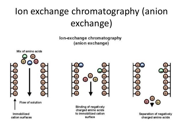


Image result for ion exchange chromatography schematic diagram
Example of anion exchange. 
Resins which have negative charges are called cation exchangers and those which have positive charges are called anion exchangers. When the solution of ions are passed through the column, the ions compete with each other for the charged sites on the resin. The rate of movement of any ion through the column depends on its affinity for the resin sites, its degree of ionization and the nature and concentration of competing ions in the solution. 

Molecules such as proteins and nucleic acids posses a variety of positively and negatively charged groups. For their separations, the differential rates of movement of ions through the column are basis for them. 

Some of anion exchangers are Dowex-2, Bio-Rad AG2, QAESephadex, Diethylam noethylcellulose. 

Some of cation exchangers are caboxymethylkellulose, BioRad AG50, Zeocarb 225. Amberlite IRC50. 

Comments

Popular posts from this blog

Statistics in Flow Cytometry Data and "MFI" values

                                 The speed of the flow cytometry offers wide range of data points and data plots. Due to its sensitivity and versatility, it has been used extensively. Flow cytometer can detect up to 1000s of cells per second.. But, it should be noted that the flow cytometry also involves the statistics, its significance, calculating the fluorescence intensity..                Statistics in flow cytometry involves, total number of data points acquired, percentage population, most importantly mean and median fluorescence intensity, and others. Most of us get confused about the term "MFI", whether it means mean fluorescence intensity or median fluorescence intensity.. Truth is, both.. It means it can be described as either mean or median fluorescence intensity. Some researchers use Mean for MFI and some use Median for MFI values.  (Keep...

Protocol: Sample preparation for Cell Cycle Analysis by Flow Cytometry

Protocol:  Preparation of Samples for Cell Cycle Analysis by Flow Cytometry: 1) Suspend approximately million cells (10^6) in 1mL of phosphate-buffered saline solution (1x). And with the help of pipette, gently aspirate about 7-8 times, to make the single cell suspension.  2) Take a centrifuge tube and add 4mL of 70% ethanol. (70% ethanol should be one ice before using). and add 1mL of cells in PBS to this tube. Keep the tubes on ice for minimum 2 hours at 4℃.. This step will fix the cells.  ( cells can kept at 4 ℃ for a week).  3) Centrifuge the ethanol fixed cells for 5 minutes at 300g. Carefully take out the supernatant. Do not disturb the pellet.  4) Add 5mL of cold 1x PBS to this pellet and suspend the cells using pipette.  5) Centrifuge the tubes at 300g for 5 minutes. and discard the supernatant. Again do not disturb the pellet.  6) if required, step 5 can be repeated for one more time.  7) Add 1mL of Propidium Iodide (PI...

Compensation in Flow Cytometry (Fluorescence Spill Over Correction).

                      The term "Compensation" in flow cytometers refers to the process of correcting fluorescence spill over. The compensation matrix ensures that the fluorescence emitted is detected in a particular detector which is from the fluorescence being measured. As we all know that most of the fluorescence molecules have broad emission range. if more than one one fluorescence is selected, their emission spectra may overlap. To correct this spectral overlap in cytometry data, a mathematical matrix called compensation is used. The Intention of this article is to teach you the compensation from basic theory to practical. This example for FITC and PE fluorescence molecules shows that, Some of the FITC fluorescence is being detected in PE Detector. and Some of the PE fluorescence is being detected in FITC Detectors. Task here is to make sure that in cytometry data, FITC is in FITC detector only and PE is in PE Detector only....