In biological sciences or bio medical sciences, the incubators are the devices which are used to maintain or grow microorganisms or cell cultures. Main task of an incubator is to maintain the optimal temperature, humidity, carbon monoxide, or CO2 and O2 content.
Incubators are regularly, in fact daily are used in clinical or research areas such microbiology, molecular biology, cell biology, etc. Commonly they are used to culture or grow bacteria as well as eukaryotic cells. Incubators are insulated boxes in which the required temperature can be adjusted depending on the cells or bacteria to be grown or cultured. The temperature can be up to 65℃ to 70℃.. Most commonly used incubators are 37℃ and 30℃ incubators. 37℃ are used for growing bacteria such as E.coli bacteria. and 30℃ are commonly used for growing yeast cells.
There are special incubators which maintain the humidity and C02 also. These conditions required for the culturing or maintaining the mammalian cells.. Commonly used C02 percentage is 5%..
There are also 4℃, 20℃, 22℃, 25℃ incubators which are called as refrigerate incubators. and here temperature can also be set according to user's convenience.
There are three main types of incubators.
1) A CO2 incubator:
usually contains 5-10% CO2 inside it and maintains constant temperature and humidity. useful for the mammalian cell culture and maintainance and tissue culture experiments. Temperature range is from 4℃ to 50℃ depending on the tissue or mammalian cells to be cultured.
2) A non CO2 incubator:
contains room air and maintains temperature which is set by the user. Commonly used for the bacterial growth and culture. some of them are used for the yeast growth also.. (But, for growing yeast,, separate incubator must be used, i.e, that incubator is meant for only yeast not for others). Temperature range is from 25℃ to 40℃.
(Example: 37℃ fro E.coli and 30℃ yeast).
3) An anaerobic incubator:
contains the atmospheric oxygen (O2) which is required to grow and maintain some cells. Used mostly for the isolation of anaerobes from the clinical samples.
Keeping in mind that these incubators come in two forms, one is static incubator in which the culture plates and others kept but not disturbed. Second on is shaker incubator in which usually culture tubes are used which rotate or shake at about 180 to 200rpm.
 |
| Integrated Scientific Solutions |
Incubators are regularly, in fact daily are used in clinical or research areas such microbiology, molecular biology, cell biology, etc. Commonly they are used to culture or grow bacteria as well as eukaryotic cells. Incubators are insulated boxes in which the required temperature can be adjusted depending on the cells or bacteria to be grown or cultured. The temperature can be up to 65℃ to 70℃.. Most commonly used incubators are 37℃ and 30℃ incubators. 37℃ are used for growing bacteria such as E.coli bacteria. and 30℃ are commonly used for growing yeast cells.
There are special incubators which maintain the humidity and C02 also. These conditions required for the culturing or maintaining the mammalian cells.. Commonly used C02 percentage is 5%..
There are also 4℃, 20℃, 22℃, 25℃ incubators which are called as refrigerate incubators. and here temperature can also be set according to user's convenience.
There are three main types of incubators.
1) A CO2 incubator:
usually contains 5-10% CO2 inside it and maintains constant temperature and humidity. useful for the mammalian cell culture and maintainance and tissue culture experiments. Temperature range is from 4℃ to 50℃ depending on the tissue or mammalian cells to be cultured.
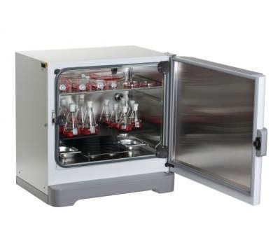 |
| New Brunswick S41i CO2 Incubator Shaker |
2) A non CO2 incubator:
contains room air and maintains temperature which is set by the user. Commonly used for the bacterial growth and culture. some of them are used for the yeast growth also.. (But, for growing yeast,, separate incubator must be used, i.e, that incubator is meant for only yeast not for others). Temperature range is from 25℃ to 40℃.
(Example: 37℃ fro E.coli and 30℃ yeast).
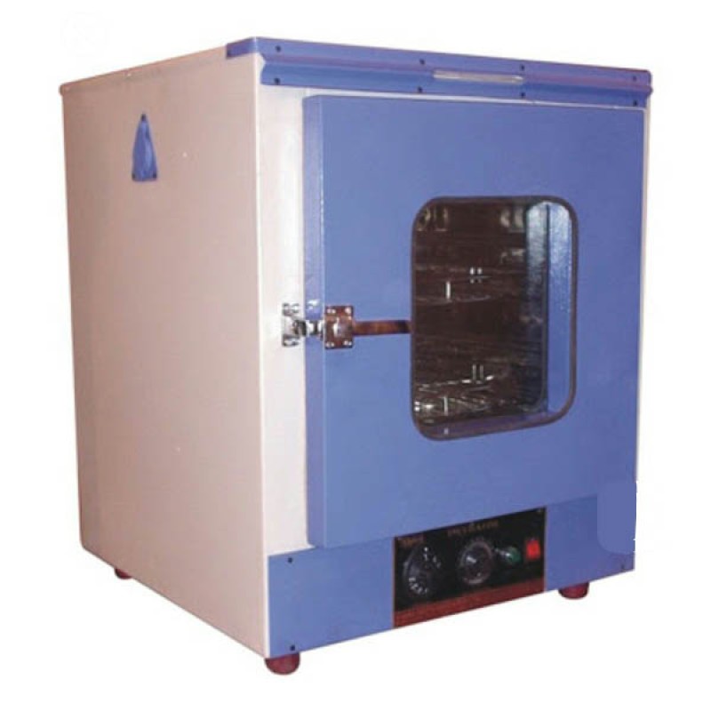 |
| Biophlox Bacteriological Incubator |
3) An anaerobic incubator:
contains the atmospheric oxygen (O2) which is required to grow and maintain some cells. Used mostly for the isolation of anaerobes from the clinical samples.
 |
| Pro-Lab Diagnostics |
Keeping in mind that these incubators come in two forms, one is static incubator in which the culture plates and others kept but not disturbed. Second on is shaker incubator in which usually culture tubes are used which rotate or shake at about 180 to 200rpm.
Comments
Post a Comment